Mastitis and types
Mastitis
Mastitis is the term which denotes inflammatory condition of udder characterized by physical, chemical and microbiological changes in milk and pathological changes in glandular tissue of the udder. In the major milk-producing countries, 15% to 20% of cows are infected with clinical mastitis each year. In Canada and the United States, it is thought that 50% of cows have one or more infected quarters.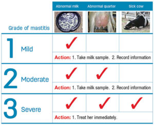
| Types of mastitis | Characteristic symptoms or definition |
|---|---|
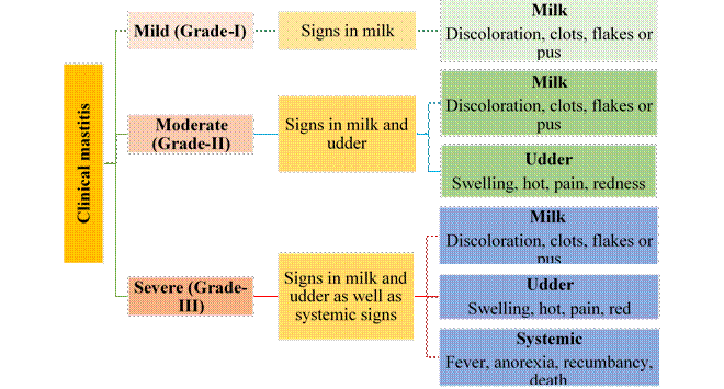
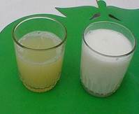
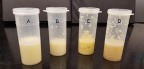
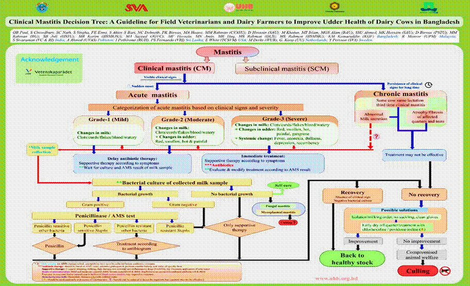
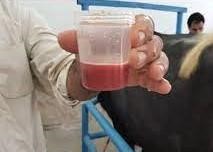
| Grade 1 (mild) | Changes to the milk only (color– off white/yellow/red and consistency– clotted/thickened) |
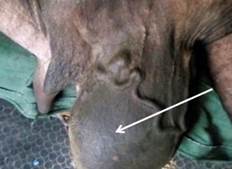
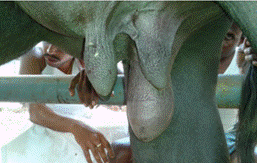
| Grade 2 (moderate) | Changes to the milk (color/consistency) and udder (heat, swelling, pain) |
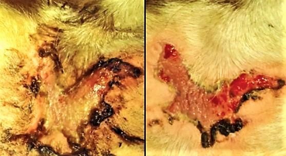
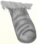
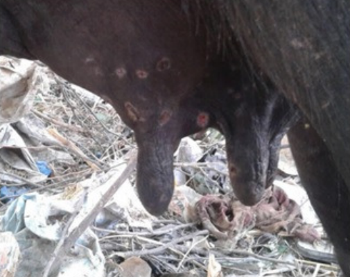
| Grade 3 (severe) | Changes to the milk (color/consistency), udder (heat, swelling, pain), cow (sick cow) and or gangrenous sloughing of the teat |
Causal agents
Microorganisms causing mastitis
There are a lot of microorganisms on and in udder of cows. 137 identified species and subspecies of microbes are identified that can be associated with the mammary gland of the cow. Several of them are part of the normal flora and, with few exceptions, do not cause mastitis. On the contrary, they may protect udders from infection caused by pathogenic bacteria. Several other microorganisms may, however, cause infection in the mammary glands. There are contagious microorganisms and environmental microorganisms. Infected cows are the main source of contagious microorganisms, which survive and proliferate on the skin and on teat wounds. They consist of Streptococcus agalactiae, Staphylococcus aureus and Streptococcus dysgalactiae. Environmental microorganisms (Escherischia coli and other coliforms, Streptococcus uberis) do not remain on the teat. Rather, their presence indicates a high degree of contamination of soil, bedding, and water caused by manure mainly.

Types of mastitis
| Species | Main Source | Living Conditions | Propagation Factors | Symptoms | Preventive Treatment |
|---|---|---|---|---|---|
| Streptococcus agalactiae | Infected cows | Infected quarter and udder only | Using same rag for cleaning udders | Mild fever for about 24 hours, Reduced milk yield, Fore strip milk having variable clots (blood stained curd) Swelling of the lower portion of udder and teat | Wash udders after milking, reduces problem by 50% Cull infected cows |
| Staphylococcus aureus | Infected cows | On abnormal udder and teat, milkers, vagina, tonsils | Transmitted by hands or rags, enters during milking | Odd colored milk containing flakes and clots, Abscess formation and infection up to deep interior of udder leading to inflammation of the upper portion of udder, Patchy blue/purple discoloration of and coldness of the affected udder In chronic state, udder hardens, aqueous secretion, eventual atrophy of the quarter. Intermediate form produces granular secretion. Milk hotter than normal | Wash udders after milking, reduces problem by 50% Cull infected cows |
| Grade 3 (severe) | Changes to the milk (color/consistency), udder (heat, swelling, pain), cow (sick cow) and or gangrenous sloughing of the teat | ||||
| Streptococcus dysgalactiae | Infected cows | Infected quarter, injuries | Reduced milk yield, Fore strip milk having variable clots (blood stained curd) Swelling of the lower portion of udder and teat High fever in serious cases | Wash udders after milking, reduces problem by 50% Cull infected cows | |
| Streptococcus uberis | Contaminated environment | On cow’s skin, mouth, ground | Neglected udder washing, insufficient drying, lack of bedding, muddy yards | Reduced milk yield, Fore strip milk having variable clots (blood stained curd) Swelling of the lower portion of udder and teat High fever in serious cases Affects mostly dry cows and heifers. | Wash teats only, dry well with disposable paper towels for each cow Supply generous bedding |
| Escherischia coli | Contaminated environment | Ground, bedding (sawdust and shavings), manure, water | Dirty calving stall, lack of bedding, inadequate udder washing | Abnormal milk (watery or blood tinge and drop in milk production, Excessive udder edema Thin yellow secretions, with granular texture resembling bran. Fever more than 106 degree F along with diarrhea and dehydration | Wash teats only, dry well with disposable paper towels for each cow Supply generous bedding |
| Corynebacterium pyrogenes | Certain insects | Humid valleys, wooded areas | Pronounced systematic reaction due to toxins caused by bacteria. Often more than one quarter affected. They become hard, produce thick smelly secretion like cheese and difficult to eliminate. Followed by abscess that bursts, releasing creamy pus, and tissue loss. | ||
| Pseudomonas | Water source, contaminated teat dips and contaminated drugs and infusion equipment, waste feed, soil and manure | Animal skin | Direct contact and using contaminated water | Marked swelling of udder, High body temperature around 107 degree F Watery milk contain flakes or clots or blood | Cull or isolate any infected cows to stop the infection from spreading, water should be tested for Pseudomonas |
| Mycoplasma | Infected animals, respiratory tracts, and urogenital tracts | Infected quarters | Cow to cow during milking, improper teat sanitation, contaminated intramammary treatments, milker’s hands, and airborne transmission | Mastitis found more than one quarter sometimes in all quarters Sharp drop in milk production leads to agalactia, Watery milk with few clots to a thick colostrum like material (Sandy or flaky sedimentation in watery or serious fluid) | Maintain a closed herd, buy replacements from known Mycoplasma-free herds, bulk tank samples may be cultured periodically |
Factors
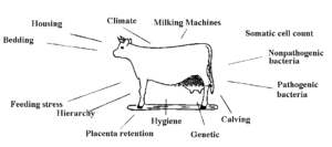
Factors contributing to mastitis
Mastitis is a difficult problem because it is a disease caused by many factors. Researchers estimate that 25% of the susceptibility to udder infection is attributable to environmental factors, 20% to genetic factors, and 50% to herd management.
Diagnosis
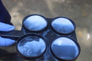
To diagnose mastitis, it is necessary to learn how to differentiate between the signs and symptoms of the various types of mastitis infection. The key points are given below:
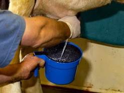
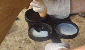
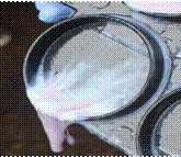
- Check the milk: Routine examination of the milk using a filter cup to extract the first three squirts before washing (before milking) is undoubtedly the best way to diagnose mastitis. The presence of lumps, flakes, blood, etc. must be watched for. Milk that is hotter than normal may be a good indication of a Staphylococcus aureus
- Palpate the udder: Particularly after milking, when it is easy to detect swelling, and fibrous, hard or injured tissue.
- Be attentive: To other more evident signs such as fever, redness etc.
If these symptoms are often absent, particularly in cases of subclinical, subacute or chronic mastitis, can be detected through observation. Some tests such as Somatic cell counts (SCC), bacterial identification and the California Mastitis Test (CMT), may be useful. These tests were discussed in diagnostic services part of the website.
Prevention
Preventive measures
Milking procedures
Sanitary milking habits are important to avoid the spreading of germs or their proliferation. The purpose of hygiene is to prevent the transmission of germs from one teat to another on one cow or from one cow to another.

Udder washing
Washing the udder is hygienic and has a stimulating effect on milk flow. Adequate washing is especially important to prevent environmental mastitis, caused by coliforms and other microbes from contaminated environments. Badly washed udders contribute to the transmission of microbes rather than to their destruction.
The lowest bacterial count in milk is obtained by washing the udder in the following way:
- Wet and wash the teats only using individual moist paper towels. Wetting the udder and the teats results in more bacteria getting into the milk than if only the teats are wet.
- Dry with individual paper towels.
Note that teat dipping before milking in addition to drying off does not give better results than drying alone, and it increases the risks of contamination of the milk by disinfectants.
Fore milking
Removing a little milk by hand before machine milking/ hand milking serves to stimulate milk letdown and to obtain a milk sample containing a high microbial count. A filter cup is used to detect abnormal looking milk (lumpy, flakes, clots, blood etc.).
Milking sequence
It is important to milk infected cows last. If possible, milking sequence should be as follows: first lactation cows, normal cows, cows with a high cell count and then infected cows.
Other measures during milking
It is important to milk completely. With modern milkers, as long as they are well adjusted, the risks of forcing the entry of microbes at the end of milking greatly diminish. The chances of bacteria entering the udder can be reduced by diminishing the amplitude of the vacuum changes and the vacuum change speed on the teats. To do so, a good vacuum reserve and appropriate piping are necessary.
Risk of infection may be diminished if milking is finished by hand, although not realistic for an entire herd. It is important to milk twice a day, even with cows that do not produce a lot. The longer the milk remains in the udder, the greater the risk of infection. The first squirts of milk must not go on the ground as this will contaminate the bedding and floor.
Postmilking teat dipping
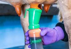
Using a disinfectant teat dip after each milking is a means of diminishing by about 50% the risk of infection by contagious microorganisms like Streptococcus agalactiae and Staphylococcus aureus. Teat dipping prevents populations of these microbes from developing sufficiently between milking. Teat dipping also discourages flies.
It is important that the teat dip contain up to 10% of emollients to increase the suppleness of the teats: oils, glycerine, lanoline. Healthy supple skin is an extra insurance against entry of bacteria to the udder. Staphylococcus aureus does not persist on healthy skin.
For more information please “Dry cow therapy” in Udder Health: Cattle part of the website
Cleaning equipment after milking
It is vital to clean and disinfect equipment after milking. Cider or corn vinegar and peroxide are used by some producers as alternatives to phosphoric acid and chlorine.
Hygiene and safety
Abundant bedding prevents injury to the udder, limits exposure to cold, damp floors and limits contact of the udder with manure. A minimum of 3 kg of straw per day per animal must be used (about one ton per cow per year). It is better to use a little bedding often, rather than a large quantity less often. Straw is preferred. Adding lime to the bedding can help in a stable where environmental mastitis is a problem but can also irritate the udder, the teats and the lungs when airborne.
It is important to keep away the cows from injuring their udders. The floors should not be slippery when the cows are let outdoors and there should be separators between the cows. The stable should be disinfected twice a year.
Feed
Changing feed must be done slowly. Excesses must be avoided, particularly concentrates and non-protein nitrogen feed. Calcium to phosphorous ratio (1.4 to 1.8) must be maintained, even during the dry period. Selenium and vitamin E supplements may be a good choice if the ration does not provide the required amount.
Replacement of stock
Farmer should avoid to infected animals for their farm. Animals have them tested before purchasing them and examine the udders carefully. Research reported that up to 50% of purchased cows have subclinical infections. It is better to buy only heifers (heifers generally do not have mastitis) or produce your own replacement animals. Heifers should not be suckled because this breaks the teat seal and thus facilitates entry of microorganisms that can cause mastitis at calving.
Culling
Cull those animals that are severely or affected by mastitis repeatedly. Cows with injured teats that do not heal should be put at the top of the list of animals to cull. They are up to 10 times more prone to contract mastitis. Cows that maintain a high cell count during all lactations should also be culled.
Drying off period
It is well known that mastitis often affects cows that have dried off recently. These animals should not be overfed.
In conventional agriculture, dry cows are treated with antibiotics along with the pre and post milking teat dip, one of the most effective methods to reduce incidence of mastitis. For organic agriculture, the dry cow must not be overlooked. A change in feed at drying-off is important. The following are the three steps:
Post-lactation (7 to 14 days): At post-lactation stage give a reduced diet of fibrous and poor hay to provoke rapid drop in milk flow and to stimulate the rumen. Drinking water must be drastically reduced. Some organic farmers give 4 drops of sage or menthol essential oil and charcoal two times a day to cut production at this stage.
Dry (30-90 days): At dry period diet is made up mostly of roughage with a good energy-protein and mineral balance.
Pre-lactation (7 to 14 days before calving): Moderate quantities of energy-rich concentrates are added to a balanced roughage ration.
Use of the teat dip before and after the dry period (i.e. 15 days before calving and 15 days after the dry period) may be beneficial in herds where clinical mastitis is common.
For more detail about dry cow therapy please visit “Dry cow therapy” in Udder Health: Cattle part of the website.
Treatment strategies
Treatment strategies for mastitis
It will depend on whether
- The mastitis is clinical or subclinical: acute or chronic
- Health status of the herd
- History of mastitis
Treatment decisions during lactation should be considered only for acute clinical cases of mastitis. Chronic cases will respond poorly to antibiotic therapy. Subclinical mastitis has a tendency to self-cure. Instead, subclinical mastitis and some chronic cases can be treated in the dry period only. (Nordic guidelines for mastitis therapy, NMSM Annual Conference, 2009)
Choice of Drugs
- Antibiotics
- NSAID (Non- steroidal anti-inflammatory drugs)
- Supportive therapy (Fluid therapy)
Indication of Antibiotics
Indication of antibiotics to treat mastitis depends on some factors:
- Type of pathogen involved

- Type and severity of inflammatory response
- Duration of infection
- Stage of lactation
- Age and pregnancy status of the cows
After taking decision of using antibiotics, decisions should be made in which route antibiotics will be administered.
Route of administration
Where to target antimicrobial therapy in clinical mastitis due to different pathogens (Pyörälä 2009, modified from Erskine 2003): Some common udder pathogens location in milk, udder parenchymal tissue or as systemic infection (number of + indicate where you mainly should target your antimicrobial therapy).
| Pathogen | In milk | In milk | In udder tissue | As systemic infection | Route of administration |
|---|---|---|---|---|---|
| Streptococcus agalactiae | +++ | - | - | Intramammary | |
| Other streptococci | +++ | + | - | Intramammary | |
| Staphylococcus aureus | + | +++ | - | Intramammary and intramuscularly | |
| Coagulase negative staphylococci | +++ | - | Intramammary | ||
| Trueperella pyogenes | - | ++ | +++ | Intramammary | |
| Coliforms* | + | - | +++ | Intramammary |
*Antibiotics are often not needed, NSAIDs and other supportive therapy is first choice.
Selection of antibiotics
Selection of antibiotics for the mastitis treatment is based on
- Culture and susceptibility testing
- About 10-40% of clinical mastitis shows no growth of bacteria, so antibiotics need not be used.
- A large proportion of Gram-negative bacteria are removed by cows own immune system, so antibiotics may be avoided.
Specific Antibiotics selection
Beta-lactamase:
Beta-lactamases are enzymes produced by bacteria that give resistance to β-lactam group of antibiotics. The major antibiotics in this group are penicillin, cephalosporin, and carbapenems. The bacteria which are capable of producing this enzyme are known as beta-lactamase-producing bacteria (BLPB). BLPB can cause multiple types of infections in humans and animals. These organisms (BPLB) indirectly release free enzyme (beta-lactam) into their surrounding environment to protect themselves & other penicillin-susceptible bacteria from penicillin therapy.
| Gram-positive bacteria, β-lactamas- | |
|---|---|
| First choice | Treatment with Penicillin G Supportive treatment. |
| Second choice | Only supportive therapy No antibiotics. |
| Gram-positive bacteria, β-lactamas+ | |
| First choice | Only supportive therapy, No antibiotics. |
| Second choice | Treatment with a β-lactamase stable antibiotic (cloxacillin, dicloxacillin, flucloxacillin, methicillin) Supportive treatment. |
| Gram-negative bacteria (E. coli) | |
| First choice | Only supportive therapy No antibiotics. |
| Second choice | Treatment with an antibiotic effective against gram-negative bacteria Supportive treatment. |
| Gram-negative bacteria (Klebsiella spp.) | |
| First choice | Treatment with med Quinolones Supportive treatment should be added. |
| Second choice | Treatment with Trimetoprim Sulfa Supportive treatment. |
| Organisms | Length |
| Staphylococcus aureus | 5 days If not cured after 5 days, the treatment could be extended for another 1 – 2 days. |
| Streptococcus uberis | 5 days |
| Other gram-positive bacteria | 3 – 5 days according to severity and herd. |
| Klebsiella spp. | 3 days |
| Other gram-negative bacteria | 3 days |
| No growth | Stop the antibiotic treatment. |
Note: Farmers should complete the full length of the treatment. They should not stop before if the cow gets better and they should not extend too long either. It will take time for the clinical symptoms to resolve even if the bacteria are gone. If the animal does not respond to the treatment with correct antibiotic and dose, the diagnosis should be confirmed at a certified mastitis laboratory as soon as possible.
Guidelines for antibiotic use for treatment of mastitis
- Milkers should be trained to detect mastitis cases early and collect milk samples aseptically. Samples should be sent to laboratory to get a basic diagnosis (no growth, Gram positive or Gram negative) to guide therapy. Cows with mild or moderate infection of clinical mastitis should be isolated and milk discarded for 24 hours until culture results are known. If the farmer wishes to initiate treatment can be given but the treatment can be modified after culture results are known
- Treatments should be administered only after a person who works closely with the local veterinarian, has reviewed the medical history of the cow and evaluated the chances for therapeutic success. Cows that are third lactation or greater, have a history of previous clinical cases, or have a history of chronically high SCC are often poor candidates for routine therapy. Treatment decisions for these cows should be based on culture results. In other instances, culling, drying off the affected quarter, or extended duration therapy may be preferred
- Only acute clinical mastitis cases should be chosen for treatment decision
- Sub-clinical mastitis in general has high self-cure or too low cure rate in proportion to the treatment costs during lactation
- Sub-clinical mastitis should be treated during the dry period
- Results of treatments should be monitored. The rate of recurrence (within 60 to 90 days) and SCC reduction (by 60 days) should be recorded
Supportive treatment
Milking frequency should be increased using the milking machine or in hand milking in cases. Milk let down can be influenced by oxytocin I/M injection. Intravenous or oral fluid therapy can be administered. 1-2ml Oxytocin injection can be administered to milk let down in cows (Oxin®, Oxitocina Diana®, Uni-Oxytocin®). Oxytocin should mainly be used for mastitis caused by Gram-negatives (can even have a negative effect on mastitis caused by streptococci). The animal can be milked after 5-10 minutes. Caution is a must during prepartum usage of oxytocin. For prepartum usage cervix must be dilated naturally or artificially by estrogen or prostaglandin. Antihistamines should be used in case of gangrenous mastitis. Anti-inflammatory drugs can be used in swollen painful udder and affected general condition or as a treatment of endotoxins released by e.g. Gram-negative pathogens. NSAIDs are mainly recommended for moderate and severe cases of mastitis. Intravenous or oral fluid therapy can be administered if the cow is dehydrated.
Prognosis evaluation:
If replacement animals are in stock, it should be used for culling as an alternative for doubtful prognosis. Cost for culling and effective treatment costs should be comparatively evaluated.
References
Akyuz A, Boyaci S, Cayli A. 2010. Determination of critical period for dairy cows using temperature humidity index. J Anim Vet Adv 9: 1824-1827.
Bargeloh, J.F. and R.O. Thomas. 1976. Relationship of mastitis and urea in rations as measured by certain milk and blood constituents. West Virginia Agriculture and Forestry, 6(3):5-7, 17.
Batra, T.R., M. Hidiroglou and M.W. Smith. 1992. Effect of vitamin E on incidence of mastitis in dairy cattle. Canadian Journal of Animal Science, 72(2):287-297.
Brim, M. and L.L. Timms. 1989. In vitro growth of environmental mastitis pathogens in various bedding materials. Journal of Dairy Science, 72(suppl. 1):14-15.
Chamberlain AT, Wilkenson JM. 1996. Feeding the dairy cow. Chalcombe Publications 90-91.
Dhakal IP, Dhakal P, Koshihara T, Nagahata H. 2007. Epidemiological and bacteriological survey of buffalo mastitis in Nepal. J Vet Med Sci 69: 1241- 1245.
Eckles CH. 1913. Dairy cattle and milk production. The Macmillan Company, New York, 342.
Eckles, C.H. 1913. Dairy cattle and milk production. MacMillan, New York. 342 pages.
Emmert, M. and K. Wendt. 1991. [Correlations between feedinq-related metabolic disorders and damage to udder health in dairy cows]. Monatshefte für Veterinärmedizin, 46(15):538-542.
Giesecke WH, Du Preez JH, Petzer IM. 1994. Practical Mastitis Control in Dairy Herds. Diagnosis of udder health problems, Butterworth Publishers: Durban, South Africa.
Giesecke, W.H. 1985. The effect of stress on udder health of dairy cows. Onderstepoort Journal of Veterinary Research, 52:175-193.
Grindal, R.J. 1988. The role of the milking machine in mastitis. British Veterinary Journal, 144:524-533.
Grohn YT, Erb HN, McCulloch CE, Saloniemei HS. 1990. Epidemiology of mammary gland disorders in multiparous Finnish Ayrshire cows. Prev Vet Med 8: 241-252.
Harmon RJ (1994) Physiology of mastitis and factors affecting somatic cell counts. J Dairy Sci 77: 2103-2112.
Hogan, J.S. , K.L. Smith, K.H. Hoblet, D.A. Todhunter, P.S. Schoenberger, W. D. Hueston, D.E. Pritchard, G.L. Bowman, L.E. Heider, B.L. Brockett, H.R. Conrad. 1989. Bacterial counts in bedding materials used on nine commercial dairies. Journal of Dairy Science, 72(1):250-258.
Jones GM. 2001. Cold weather mastitis prevention tips. Dairy Pipeline.
Jones GM. 2009. The Role of Milking Equipment in Mastitis. Virginia Cooperative Extension.
Keller, P. 1977. The influence of the environment on the health of cows in cubicle stalls. Proceedings of a seminar on Agricultural Buildings, As, Norvège, Section II, pages 118 to 124.
Klastrup, O., G. Bakken, J. Bramley and R. Bushnell. 1987. Environmental influences on bovine mastitis. Bulletin of the international dairy federation, No. 217, 37 pages.
Klug, F., H. Franz, B. Bethge, G. Jansch, F. Lemme. 1989. [Effects of level of nutrition during early lactation on health and conception rate of group-fed dairy cowsl. Tierzucht, 43(2):56-57.
MacLeod, G. 1981. The treatment of cattle by homeopathy. Health Science Press, Saffron Walden, Essex, England. 148 pages.
Mein GA, Neijenhuis F, Morgan WF, Reinemann DJ, Hillerton JE, et al. 2001. Evaluation of bovine teat condition in commercial dairy herds: 1. Non-infectious factors. Proceedings of the 2nd International Symposium on Mastitis and Milk Quality, NMC/AABP, Vancouver, 374-351.
Merck, C.C., B. Sonnenwald and H. Rollwage. 1989. [Studies in the treatment of acute bovine mastitis with homeopathic drugsl. Berliner und Munchener Tierarztliche Wachenschrift, 102(8) :266-272.
Milojevic, Z., M. Siradovic, D. Marovic, D. Sandor, R. Micic, S. Kojevic, M. Ismailovic and S. Filipovic. 1988. [Effect of various management systems on udder infections and the occurrence of mastitis]. Nauka u Praski, 18(2):231-236.
Morse, D., M.A. Lorenzo, C.J. Wilcox, R.J. Collier, R.P. Natzko, D.R. Bray. 1988. Climatic effects on occurrence of clinical mastitis. Journal of Dairy Science, 71 (3):848-853.
Ndiweni, N. and J.M. Finch. 1991. The relationship between vitamin E-selenium status and the incidence of mastitis in dairy herds near Harare. Zimbabwe Veterinary Journal, 22(4):101 -109.
Newman LE, Kowalski JJ. 1973. Fresh Sawdust Bedding-A Possible Source of Klebsiella Organisms. Am J Vet Res 34(7).
Nordic guidelines for mastitis therapy, NMSM Annual Conference, 2009
Oliver, J., F.H. Dodd and F.K. Neave. 1956. Udder infections in the dry period. 5. The effect of teat disinfection at drying-off on the incidence of infections in the early dry period. Journal of Dairy Research, 23:212-216.
Pankey, J.W. 1989. Hygiene at milking time in the prevention of bovine mastitis. British Veterinary Journal, 145:401 -409.
Philpot, W.N. 1978. Prevention of mastitis by hygiene. pages 547 to 562. In Wilcox, C.J. et al.. 1978. Large dairy herd management. University of Florida, Gainesville, Florida. 1046 pages.
Philpot, W.N. and F.H. Dodd. 1978. Mastitis. Chapter 23 In Wilcox, C.J. et al.. 1978. Large dairy herd management. University of Florida, Gainesville, Florida. 1046 pages.
Pouden, W.D., J.W. Hibbs and B.H. Edging on. 1952. The activity of streptococcus agalactiae in milk possibly influenced by the ration. American Journal of Veterinary Research, 13:486-499.
Quiquandon, H., 1982. Veterinary Medicine and Organic Farming. Biotherapic Medicine in Livestock. In Basic Technics in Ecological Farming / Basic Techniques in Organic Farming / Grundsätzliche Verfahren der ökologischen Landwirtschaft / The Maintenance of Soil Fertility / The Maintenance of Soil Fertility / Die Erhaltung der Bodenfruchtbarkeit (pp. 149-170
Radostits, O.M. 1961. Coliform mastitis in cattle. Canadian Veterinary Journal, 2:201-206.
Ranjan R, Gupta MK, Singh KK. 2011. Study of bovine mastitis in different climatic conditions in Jharkhand, India. Vet World4: 205-208.
Schukken YH, Grommers FJ, Van de Geer D, Brand A. 1989. Incidence of clinical mastitis on farms with low somatic cell counts in bulk milk. Vet Rec 125: 60-63.
Schukken YH, Grommers FJ, Van de Geer D, Brand A. 1989. Incidence of clinical mastitis on farms with low somatic cell counts in bulk milk. Vet Rec 125: 60-63.
Schukken, Y.H., H.N. Erb and J.M. Scarlett. 1989. A hospital-based study of the relationship between retained placenta and mastitis in dairy cows. Cornell Veterinarian, 79(4):319-326.
Sentitula, Yadav BR, Kumar R. 2012. Incidence of Staphylococci and Streptococci during winter in mastitic milk of sahiwal cow and murrah buffaloes. Ind J Microbiol 52: 153-159.
Seykora AJ, McDaniel BT. 1985. Udder and teat morphology related to mastitis resistance: a review. J Dairy Sci 68: 2087-2093.
Shaldon JP. 1980. Dairy farming: being the theory, practice, and methods of dairying (1880). Cassell and Company, London 575.
Shathele MS. 2009. Weather effect on bacterial mastitis in dairy cows. Intl J Dairy Sci 4: 57-66.
Smith KL and Hogan JS. 2000. Bedding’s Contribution to Mastitis in Dairy Cows. Dairy Housing and Equipment Systems, Managing and Planning for Profitability. NRAES 129.
Smith, K.L., J.S. Hogan and B.P. Weiss. 1989. Dietary selenium and vitamin E influence the resistance of cows to mastitis. Pages 27 to 32 In Proceedings of the British Mastitis Conference. 1989. The environment and mastitis. Cambridge, UK.
Sordillo LM, Shafer-Weaver K, DeRosa D. 1997. Immunobiology of the mammary gland. J Dairy Sci 80: 1851-1865.
Sterk, V., R. Beslin, A. Anojcic and A. Pavlicevic. 1978. [Effect of method of feeding on the defence capacity of the udder in dairy cows]. Veterinarski Glasnik, 32(11):899-903.
Stowell RR, Inglis S, 2000. Sand For Bedding. Dairy Housing and Equipment Systems. NRAES 129.
Tiwari JG, Babra C, Tiwari HK, Williams V, Wet SD, et al. 2013. Trends In Therapeutic and Prevention Strategies for Management of Bovine Mastitis: An Overview. J Vaccines Vaccin 4: 176. doi:10.4172/2157-7560.1000176
Underwood EJ, Suttle NF. 1999. In: The Mineral Nutrition of Livestock. Underwood EJ and Suttle NF (eds), CABI Publishing, New York.
Upadhayay AK, Gangwar P, Kumar M. 2008. Supplementation to prevent subclinical mastitis. Vet World 1: 40-41.
Weiss, W.P., J.S. Hogan, K.L. Smith and K.H. Hoblet. 1990. Relationships among Se, vitamin E and mammary gland health in commercial dairy herds. Journal of Dairy Science, 73(2):381-390.
Whittaker, J. 1995. Seeking the nutrition factor in mastitis. Acres USA, 15(11):41.
www.printfriendly.com (Responsible Use of Antibiotics for Treatment of Clinical
www.progressivedairy.com/topics/management/make-better-decisions-with-mastitis-grading-chart
www.svenskmjolk.se (Nordic Guidelines for Mastitis Therapy)
Zehner MM, Farnsworth RJ, Appleman RD, Larntz K, Springer JA. 1986. Growth of Environmental Pathogens in Various Bedding Materials. J. Dairy Sci. 69(7).
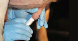

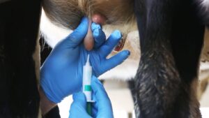 t sealant and may be especially valuable for the longer dry period. Using the internal sealant in combination with an antibiotic prevents significantly more new dry period infections than using antibiotic alone.
History
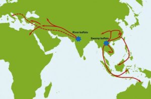
The domesticated water buffalo is a potential livestock species that supplies humankind with milk, meat, and draught power. This species is reared in almost 77 countries covering Asia, Europe, Africa, and America. Two types of domestic Asian water buffalo descended from different wild Asian water buffalo (Bubalus arnee) populations some 900 thousand years ago which later spread into other geographical regions. It is believed that the river buffalo (B. bubalis bubalis) was domesticated in the western region of the Indian subcontinent (6300 years ago) and spread to Egypt, the Balkans, Greece, and Italy. On the other hand, swamp-type (B. bubalis carabanesis) buffalo were domesticated near the border of the China and Indochina region 3000-7000 years ago and dispersed through Southeast Asia, Assam and Bangladesh, and China
Distribution
Because of the high-quality products and their adaptability to harsh environmental conditions, interest in buffalo production has increased. Buffaloes are found in India, Pakistan, Bangladesh, Iraq, Iran, Nepal, Myanmar, the Philippines, Turkey, Italy, China, Thailand, Brazil, and Egypt. The number of buffaloes worldwide was reported to be 206 million in 2018. Asia has the majority of the buffaloes (97.3%), followed by Africa (1.7%), America (0.7%), Europe (0.2%), and Oceania (0%). Table-1 shows that India has the largest buffalo population of all countries. This buffalo population contributes 55% of the total milk in the country. Some of the best and most productive buffalo breeds originated from India, such as Murrah, Nili-Ravi, Surti, and Jafarabadi.
Buffalo population in different countries
| Countries | Buffalo population (in million) |
| India | 114.15 |
| Pakistan | 38.84 |
| China | 27.11 |
| Brazil | 1.39 |
| Italy | 0.40 |
| Malaysia | 0.11 |
| Sri Lanka | 0.30 |
| Thailand | 1.25 |
| Vietnam | 2.42 |
| Philippines | 2.88 |
| Myanmar | 3.79 |
| Nepal | 5.27 |
| Bangladesh | 1.48 |
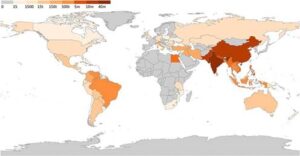
Figure 1.2: World buffalo distribution map (Credit: Minervino et al., 2020)
Types of buffalo breeds
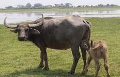
The two major types of wild buffaloes are Asian water buffalo (Bubalus arnee) and African Buffalo (Syncerus caffer). The two subspecies of Asian water buffalo are: the river buffalo (B. bubalis bubalis), most found in the Indian subcontinent and some European and American countries, and the swamp type buffalo (B. bubalis carabanesis,) mainly found in Bangladesh, China, Southeast Asia, Australia, and north-eastern states of India.
| Points | River type buffalo | Swamp type buffalo |
| Distribution | India, Pakistan, Bangladesh, some European and American countries | Bangladesh, China, Thailand, the Philippines, Indonesia, Australia, the North-eastern states of India, etc. |
| Chromosome no. | 50 chromosomes | 48 chromosomes |
| Horn | Relatively straight pale-colored horns or curled. | Generally curved, massive backswept horns. |
| Color | Black or dark grey without white markings on the leg and face, mainly | Slate gray with white chevron (one or two white stripes on the throat), socks, and tail tip. |
| Adult weight | 450 - 1000 kg | 325 - 450 kg |
| Production | Milk production is higher than swamp-type buffalo | The females yield up to 600 kg of milk per lactation |
| Utility | Mainly for milk production and suitable for meat and draught purposes as well | They are mainly used for draught purposes and are poor milk producers. |
| Figure |  |
 |
Some important buffalo breeds of Asia and Europe are-
| Traits | Picture |
| Murrah
● Origin: The center of Haryana and spread across the country. ● Distribution: Different states of India, Brazil, Bulgaria, and many Asian countries. ● Skin and hair color: Jet black with a white switch in the tail. ● Horn: Short and tightly curled. ● Body weight: o Bull: 750 kg o Cow: 650 kg ● Milk production: 1,800 kg ● Lactation period: 305 days ● Milk fat: 7.2% |
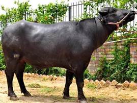
Fig: Murrah Bull |
| Nili-Ravi
● Origin: Around the Ravi River, India. ● Distribution: Ferozpur district, Punjab, and Sahiwal (Pakistan). ● Body color: Black with white spots in extremities and walled eyes. ● Horns are less curled compared to Murrah buffalo. ● Udder: Well-shaped and extends well forward to the naval flaps. ● Body weight: o Bull: 700 kg o Cow: 600 kg ● Milk production: 2,000 kg ● Lactation period: 305 days ● Milk fat: 6.5% |
 Fig: Nili-Ravi bull(Credit: Hisarbovine) |
| Kundi
● Origin: Indus Valley ● Distribution: Sindh region of south Pakistan ● Body color: Jet black ● The horn is short with a broad base and tapers upward and inward ● Udder is well developed with prominent milk veins, and teats are squarely placed ● Body weight: o Bull: 700 kg o Cow: 600 kg ● Milk production: 2,000 kg ● Lactation period: 320 days ● Milk fat: 7% |
 Fig: Kundi |
| Surti
● Origin: Southwestern part of Gujarat ● Distribution: Gujarat and Rajasthan district of India. ● Black coat color with black or reddish skin and two white chevrons on the chest ● Characterized by white markings on the forehead, legs, and tail tips. ● Horns are flat, medium length, and directed downward and backward ● Body weight: o Bull:700 kg o Cow: 550-650 kg ● Milk production: 2,090 kg ● Lactation period: 350 days ● Milk fat: 6.6-8.1% |
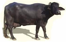 Fig: Surti (Credit: TradeIndia) |
| Jafarabadi
● Origin: Gujarat, India. ● The breeding tract is Gir forests, Kutch, and Jamnagar districts of Gujarat in India. ● Amber-black in color with white or gray marking forehead, tail, and feet ● Horn is broad and large, directed downward and then upward. Sometimes covers the eyes. ● Body weight: o Bull: 600-1,500 kg o Cow: 700-800 kg ● Milk production: 1,800-2,700 kg ● Lactation period: 350 days ● Milk fat: 8.5% |
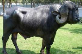 Fig: Jafarabadi (Credit: ExportersIndia) |
| Mediterranean Buffalo
● Origin: IndiaInvalid source specified. ● Mainly distributed throughout Italy ● Medium body with grey hair ● Mostly black, black and brown, and dark gray ● The horn is flat at the bottom and faces backward, and their points face upward and inward ● Body weight: o Bull: 500-600 kg o Cow: 300-450 kg ● Milk production: 900-4,000 kg ● Lactation period: 270 days ● Milk fat: 8% |
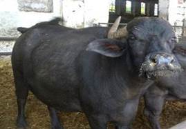 Fig: Mediterranean Buffalo |
| Indigenous Bangladeshi
● Found mainly in the Southwestern part of Bangladesh. ● Medium size body and black with a white spot on the forehead and tail-switch. ● The horn is medium to large and directed backward to outward. ● Body weight: 427 kg ● Milk production: 620 kg ● Milk fat: 6.8-13.2% |
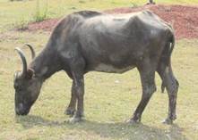 Fig: Indigenous Bangladeshi(Photo credit: Sanjib Chandra Nath) |
Behaviour
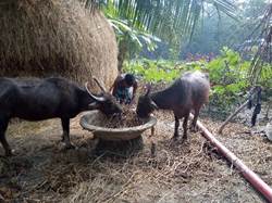
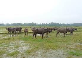
Feeding behaviour: Buffaloes are strict grazers and usually graze during the daytime, while ruminating and sleeping are predominant activities at night. They are good converters of poor-quality roughage to high energy. These animals spend more time in rumination, less time in ingestion, and more at rest than cattle.
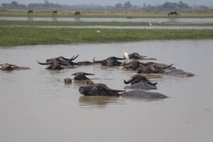
Heat tolerance and wallowing behaviour: Buffaloes are less tolerant to extreme heat and cold than cattle. They have thick skin with many melanin pigments, giving a black coat color. This thick and black coat color is a good heat absorber and limits heat loss from the body. Moreover, a lower density of sweat glands and hair in the skin limits heat loss. As a result, buffalo show wallowing behaviour, which is a learned behaviour. Buffaloes wallow in water during hot and cold weather. It helps to regulate their body temperature and reduce heat stress. In another way, wallowing is helpful to protect them from insects.
Social behaviour: Like many other livestock species, buffaloes live in groups of bulls, dry or pregnant cows, young stock, and calves. They communicate through different visual signals, sound, touch, and smell. The buffaloes recognize each other by smelling each other. Buffaloes will rub themselves against trees to leave a scent and defecate to mark their territory. After parturition, the dam stands up and starts to lick and sniff her calf to stimulate respiration, blood circulation, urination, and defecation. Dams quickly learn to recognize their calf and usually reject neonates from other mothers if they try to approach their udder. Both the dam and calf use vocal communication to recognize each other.
Utility of buffalo
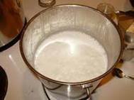
Milk production: Buffalo is the world's second-highest milk-producing animal. But in some South Asian countries like India, Pakistan, and Nepal, the contribution of buffalo milk to total milk production is higher than cow milk. Buffalo milk has higher nutritional value, for example, more protein, vitamin, and mineral content than cow's milk. Moreover, it contains beneficial compounds that may provide antioxidant protection and improve bone and heart health. The high milk solids and fat in buffalo milk aid in preparing dairy products such as yogurt and cheese.
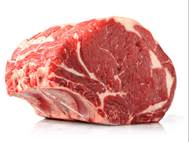
Meat production: Buffalo meat is very popular in most countries. About 90.5% of world buffalo meat was produced in Asia, with the more incredible amount contributed by India, with 1.6 million tons produced, followed by Pakistan with 0.93 million tons. Buffalo meat is known in various names in different countries. In some places, it is known as red beef, or buff in India and Nepal; in some countries, it is known as carabeef. It is lean and rich in protein but lower in fat, cholesterol, and calories than cattle meat. This unique nutritional profile has created a high demand among health-conscious consumers.
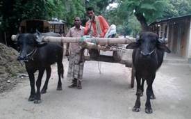
Draught purpose: For centuries, buffaloes have been used as draught animals as they have good muscular development. They are widely used to plough, puddle rice fields, level land, cultivate field crops, haul carts, sleds, shallow-draft boats, etc. The strong large feet, legs, and powerful quarters enable them to maintain balanced traction. They also carry people, thresh grain, press sugar cane, haul logs, and more. Buffaloes have an advantage over other draught animals with large hooves in wet or muddy areas. Their legs can withstand wet conditions better than cattle.
References
- N. Mingala, M. A. Villanueva and L. C. Cruz, "River and Swamp Buffaloes: History, Distribution and their Characteristics," in The Buffalo (Bubalus bubalis) - Production and Research, Bentham Science Publishers, 2017, pp. 3-31.
- Zhang, L. Colli and J. Barker, "Asian water buffalo: domestication, history and genetics," Animal Genetics, vol. 51, 2020.
- Naveena, M. Kiran, R. Banerjee and M. Muthukumar, "Water buffalo," Elsevier Ltd., 2022, pp. 1-20.
- FAO, "FAOSTAT 2018," 2018. [Online].
- Mathivanan, "Breeds of Buffaloes," TNAU, 2014. [Online]. Available: https://agritech.tnau.ac.in/animal_husbandry/animhus_buffalo%20breeds.html.
- Z. M. Minervino, D. Vecchio and B. Antonio, "Bubalus bubalis: A Short Story," Frontiers in Veterinary Science, vol. 7, 2020.
- WR, "The water buffalo: a review," British Veterinary Journal, vol. 137, p. 8–10, 1981.
- Wanapat, K. Sommart, C. Wachirapakorn, S. Uriyapongson and C. Wattanachant, "Recent advances in swamp buffalo nutrition and feeding," in 1st Asian Buffalo Association Congress, Khon Kaen University, Khon Kaen, 1994.
- Bilal, M. Suleman and A. Raziq, "Buffalo: black gold of Pakistan," Livestock research for rural development, vol. 18, no. 9, pp. 140-151, 2006.
- Singh, "The Beef About Buff," Outlook (India), 2016. [Online]. Available: https://www.outlookindia.com/magazine/story/the-beef-about-buff/297825/.
- Hill, Cattle and Buffalo Meat Production in the Tropics, Longman Scientific & Technical, 1988.